
Breast cancer surgery.
Three out of four women diagnosed with breast cancer choose breast-conserving surgery. Whether the surgeon removed all of the cancer is decided days later, by pathology. LUCY is designed to answer that question in the operating room.
A positive margin means a second operation.
1. Metcalfe et al., JAMA Surg 2017;152(11):1084.
During breast-conserving surgery the surgeon removes the tumor with a rim of surrounding tissue and sends it to pathology. The lab checks whether cancer reaches the inked edge of the specimen. That answer takes five to ten days. If the margin is positive, the patient comes back for another operation, with the anxiety, the risk and the cost that brings.
The tools available in the operating room today each fall short. Frozen section and specimen radiography sample only part of the surface. A radiofrequency probe samples points by contact. A fluorescent dye has to be injected hours before surgery.
The map appears while the patient is still on the table.
LUCY sits on a cart beside the surgical field and images the whole specimen surface from the tissue's own fluorescence. No dye, no contact probe.
- 1
Scan
The lumpectomy specimen goes into the tray and the drawer closes. One button starts the scan.
- 2
See
In under two minutes a color-coded map shows where the surface carries the signature of cancer.
- 3
Decide
The surgeon takes an additional shave from the flagged location in the same session, or closes.
- 4
Close
The aim is a complete resection in the first operation.
The specimen, the map and the pathology, side by side.
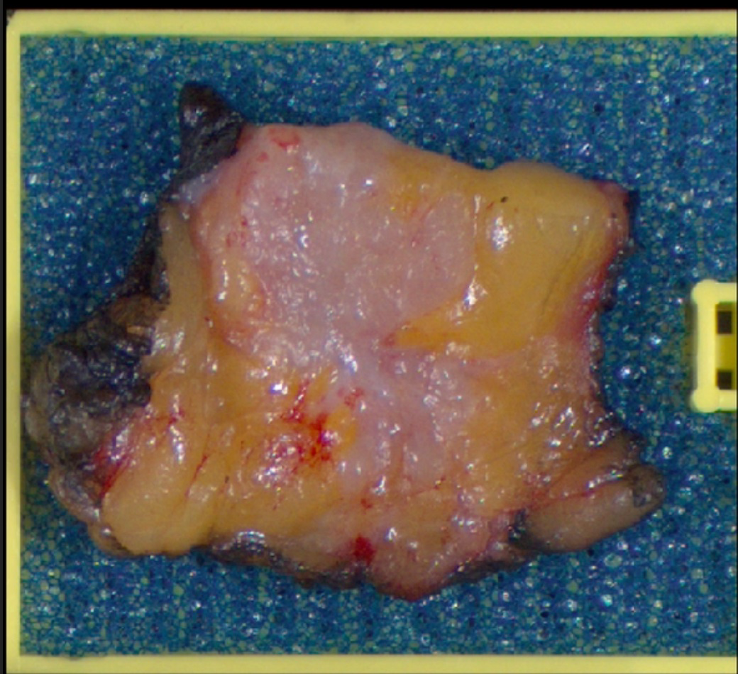 Excised tissue
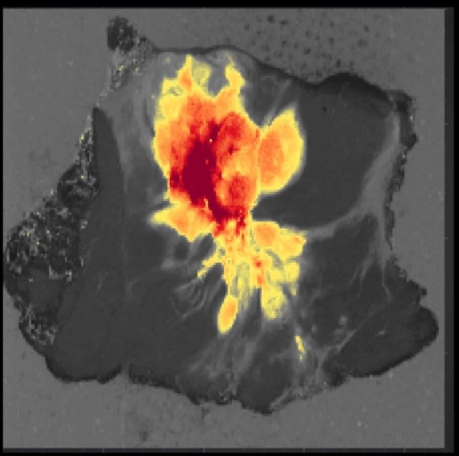 LUCY scan
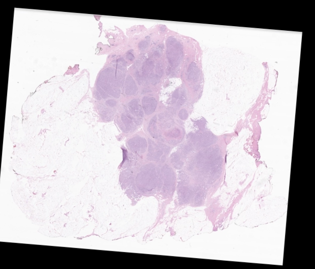 Matched pathology
Clinical studies at Cedars-Sinai, and a pivotal trial designed with FDA.
The studies imaged freshly excised human breast tissue and compared every scan with the pathology read of the same slice.
Investigational device Study results are from ex vivo clinical research and are not a substitute for FDA approval or clearance.